- #Nih imagej software function mac os x#
- #Nih imagej software function software#
- #Nih imagej software function download#
Although the look is slightly different, the overall feature.
#Nih imagej software function software#
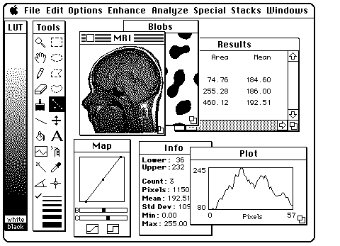
IPLaminator is an easy to use software application that will greatly speed and standardize quantification of neuron organization. (a,b) Screenshots of NIH Image 1.62, released in 1999 (a), and ImageJ 1.45, released in 2011 (b). Statistical analysis of the output then allows a quantitative value to be assigned to differences in laminar patterning observed in different models, genotypes or across developmental time. Options to analyze tissues such as cortex were also added. A range of user options allows researchers to bin IPL stratification based on fixed points, such as the neurites of cholinergic amacrine cells, or to define a number of bins into which the IPL will be divided. The novel ImageJ based software plugin we developed: IPLaminator, rapidly analyzes neurite stratification patterns in the retina and other neural tissues. One of the great things about the software is that it works with all operating systems: Mac, Linux and Windows. ImageJ software is used for image analysis and processing. It was written and is maintained by Wayne Rasband of NIH.
#Nih imagej software function mac os x#
ImageJ is written in Java, which allows it to run on Linux, Mac OS X and Windows, in both 32-bit and 64-bit modes.

#Nih imagej software function download#
In this study we report the development of an intuitive platform to rapidly and reproducibly assay IPL lamination. ImageJ is a Java based software program available as freeware for download from the ImageJ homepage. ImageJ is a free and very capable image analysis software made available from the National Institutes of Health (NIH). Most previous work on neuron stratification in the IPL is qualitative and descriptive. A current limitation to such analysis is the lack of standardized tools to quantitatively analyze this complex structure. Here we describe an open source plugin for ImageJ called EzColocalization to visualize and measure colocalization in microscopy images. Enter the email address you signed up with and well email you a reset link. Understand the capabilities of ImageJ and how this software can be used to learn about and perform image analyses. Insight into the function and regulation of biological molecules can often be obtained by determining which cell structures and other molecules they localize with (i.e. Characterization of valvular interstitial cell function in three dimensional matrix metalloproteinase degradable PEG hydrogels. Studies focused on developmental organization and cell morphology often use this layered stratification to characterize cells and identify the function of genes in development of the retina. We also do software evaluation for GE Medical Systems None of the software discussed is FDA approved for clinical use I don’t program C or Java, for that matter Objectives 1. The neurites of the retinal ganglion, amacrine and bipolar cell subtypes that form synapses in the IPL are precisely organized in highly refined strata within the IPL. This is perhaps best exemplified in the layering, or lamination, of the retinal inner plexiform layer (IPL). .ImageJ Software is open source and available to download at National Institute of Healths (NIH) website: Video was made at Ontario Tech University. Information in the brain is often segregated into spatially organized layers that reflect the function of the embedded circuits.


 0 kommentar(er)
